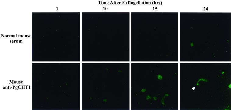
FIG. 1.
Immunofluorescence localization of P. gallinaceum chitinase PgCHT1 in in vitro-developed mosquito midgut stage parasites. Parasites were stained at the indicated time points with either normal mouse serum or anti-P. gallinaceum chitinase (PgCHT1) antibodies and visualized with FITC-labeled goat anti-mouse antibody. The white arrowhead indicates the apical end of the mature ookinete.

