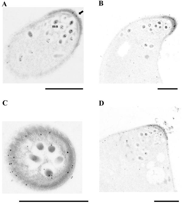
FIG. 3.
Immunoelectron microscopy of in vitro-developed mosquito midgut stage P. gallinaceum parasites stained with anti-P. gallinaceum chitinase (PgCHT1) antibodies. (A) Longitudinal section of a maturing ookinete in which PgCHT1 is associated with micronemes (mn) at the apical third of the parasite (arrow). The primary antibody was mouse polyclonal antiserum raised to full-length recombinant PgCHT1. (B) Longitudinal section of a mature ookinete stained with anti-PgCHT1 chitin-binding domain antiserum. (C) Cross section of the apical end of a mature ookinete stained with anti-PgCHT1, anti-active-site serum. (D) Longitudinal section of a mature ookinete stained with anti-PgCHT1 chitin-binding domain serum showing extracellular PgCHT1. Bars, 1 μm.

