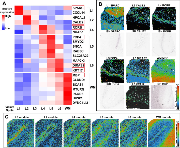
Fig. 2.
Layer-specific genes define the anatomical architecture of the human MTG and the frontal cortex. (A) Heatmap of Z-scores for layer-specific marker genes identified from CT and AD human MTG. The heatmap shows the expression levels of layer-specific marker genes in each layer except layer III, which does not show any conserved marker genes in all 6 samples. The red color indicates a relevantly higher expression than other layers, while the blue color indicates a lower expression. Identified layer-specific marker genes, SPARC (L1: layer I), CALB2 (L2: layer II), RORB (L4: layer IV), PCP4 (L5: layer V), DIRAS2 and KRT17 (L6: layer VI), and MBP (the WM), are boxed in red and were used for validation of cortical laminae in spatial maps using Loupe Browser shown in (B). (B) Spatial maps of layer-specific marker genes in our samples (color images) were validated by in situ hybridization (ISH) data (gray images) from the Allen Brain Institute’s Human Brain Atlas (Sample ID: 79,205,802, 80,225,788). ISH data is not available for DIRAS2 and KRT17. (C) Public available 10 × Visium SRT data of the human frontal cortex (Sample ID: Human Brian 1) is annotated by our layer-specific marker gene modules

