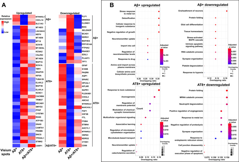
Fig. 6.
AD pathology-associated gene signatures and pathways. (A) Heatmap of Z-scores for upregulated and downregulated DEGs specific to Visium spots localized with different AD pathologies (Aβ+, AT8+, and Aβ/AT8+). DEGs were identified from pathological regions (either Aβ+, AT8+, and Aβ/AT8+) vs. surrounding level 3 spots of three AD samples at the pseudo-bulk level. The red color indicates a relevantly higher expression than other pathologies, while the blue color indicates a lower expression. (B) Gene ontology (GO) pathway analysis of identified DEGs (Additional file 10: Table S9) specific to Visium spots localized with Aβ plaques (top row) or pathological tau, AT8 (bottom row). The Dot plot shows top 10 enriched GO terms. Each dot is colored by the Benjamini–Hochberg adjusted p-value. The dot size is scaled by the number of overlapping genes with the related GO terms. The x-axis is the ratio and indicating the proportion of overlapping genes between the query gene list and all genes in the GO term

