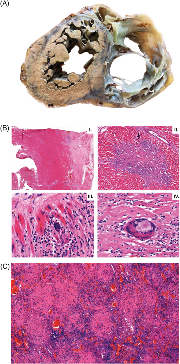
Figure 4.

Macroscopic appearance of the heart and histological findings in the heart and a mediastinal lymph node during autopsy. (A) Photograph of a transverse slice of the heart at midventricular level with extremely thin right ventricular wall. The right ventricular myocardium is mostly replaced by fibrotic tissue. The appearance is similar to arrhytmogenic right ventricular cardiomyopathy. (B) Left ventricular tissues showing fibrosis advancing from the epicardium towards the endocardium (I.). There is non‐necrotizing granulomatous inflammation (II.) with epithelioid histiocytes (III.) and multinucleated giant cells (IV.) supporting the diagnosis of sarcoidosis. (C) The histology of a firm mediastinal lymph node shows non‐necrotizing granuloma‐like structures with a few giant cells. Mycobacteria were not confirmed by Ziehl–Neelsen staining, excluding tuberculosis.
