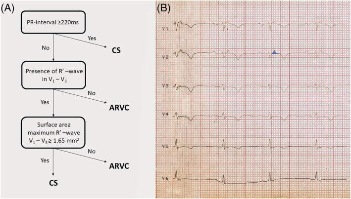
Figure 5.

The algorithm devised to distinguish sarcoidosis with left and right ventricular involvement from ACM and its application on our patient's ECG. (A) The ECG algorithm. (B) The application of the algorithm on our patient's ECG recorded after the first admission. The surface area of the maximum R′ wave in lead V2 marked by light blue colour was ≥1.65 mm2. R′ wave was defined as any positive deflection after an S wave. ARVC, arrhythmogenic right ventricular cardiomyopathy; CS, cardiac sarcoidosis. For further explanation, see text.
