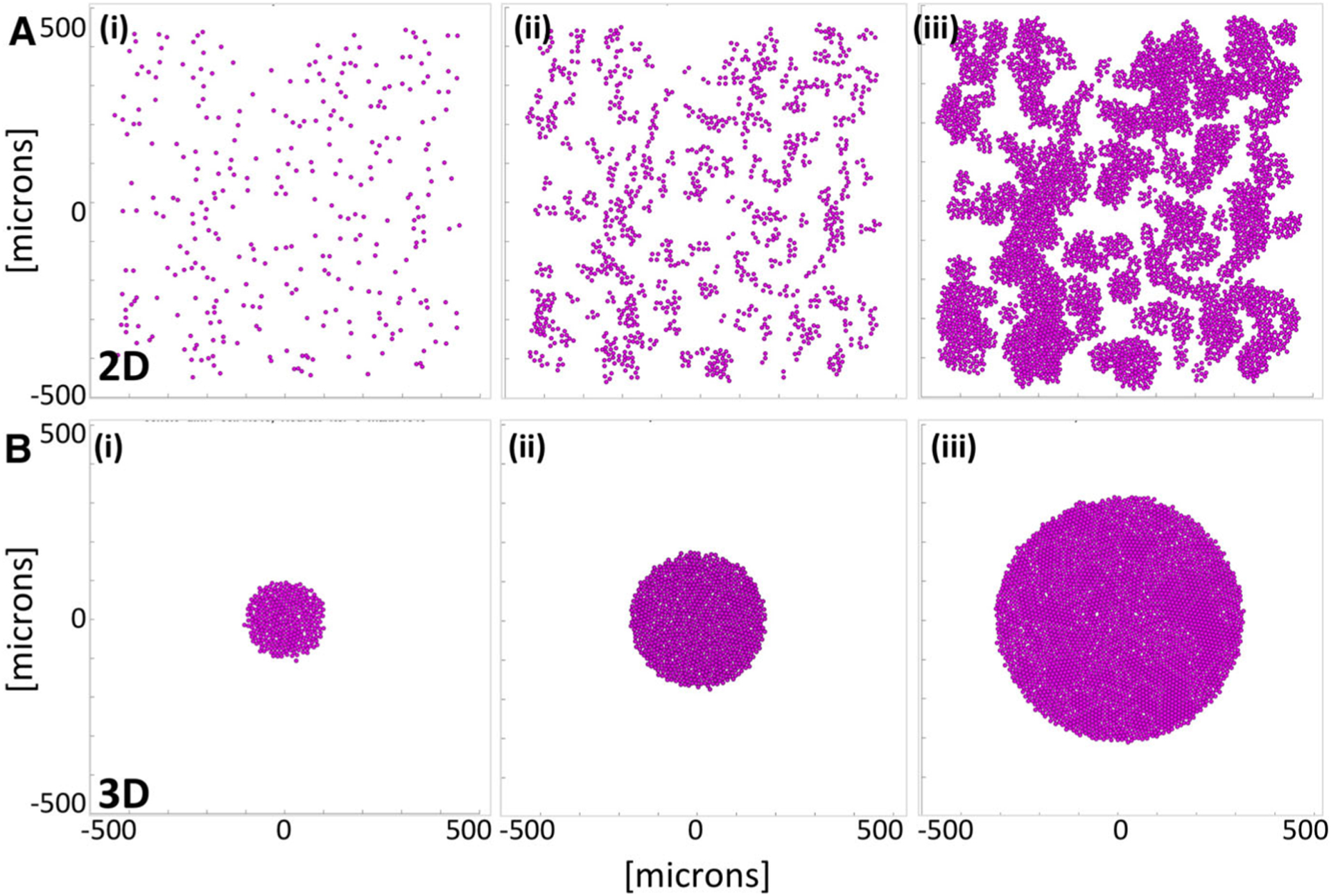
Fig. 2.

Tumor growth in 2D and 3D cell cultures with no drug (the control case). a A computational model of the monolayer cell culture with sparsely seeded initial 315 cells (i), the daughter cells divide and spread throughout the domain (ii) for 72 h of the simulated time reaching 4516 cells (iii). b A computational model of the cross section though the spheroid cell culture with initial cluster of 315 cells (i), the non-overcrowded cells divide and expand (ii) for 72 h of the simulated time reaching 3183 cells (iii)
