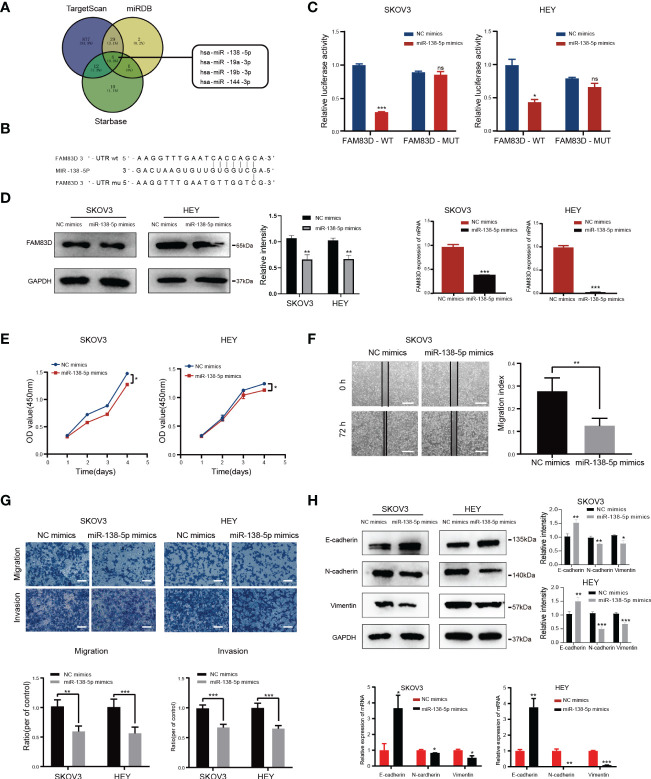
Figure 6.
FAM83D is a novel target of miR-138-5p in OC cells. (A) Bioinformatics analysis predicted that FAM83D was the target of miR-138-5p. (B) Schematic representation of the FAM83D 3’ -UTR containing the binding site for miR-138-5p. (C) The dual luciferase reporter assay confirmed that FAM83D is the direct target gene of miR-138-5p. ns, no significance, *P < 0.05, **P < 0.01 and ***P < 0.001 vs. cells transfected with NC mimics cells (t-test, N = 3) (D) Western Blot and qRT-PCR analysis of FAM83D expression in SKOV3 and HEY cells transfected with miR-138-5p mimics or NC mimics for 48 h The protein levels were quantified by grey analysis and showed in the right panel. **P < 0.01 and ***P < 0.001 vs. cells transfected with NC mimics cells (t-test, N = 3). (E) Growth curve of HEY and SKOV3 cells upon transfection with NC mimics and miR-138-5p mimics examined by CCK8 assay. *P < 0.05 vs. cells transfected with NC mimics cells (t-test, N = 3). (F) Scratch wound healing showed the migratory capacities of HEY and SKOV3 cells upon transfection with NC mimics and miR-138-5p mimics. Scale bar: 500 μm. **P < 0.01 vs. cells transfected with NC mimics cells (t-test, N = 3). (G) Cell migration and invasion ability were measured by Transwell assays. Scale bar: 200 μm. **P < 0.01 and ***P < 0.001 vs. cells transfected with NC mimics cells (t-test, N = 3). (H) Western blot and qRT-PCR analysis of the expression of EMT markers. The protein levels were quantified by grey analysis and showed in the right panel. *P < 0.05, **P < 0.01 and ***P < 0.001 vs. cells transfected with NC mimics cells (t-test, N = 3).

