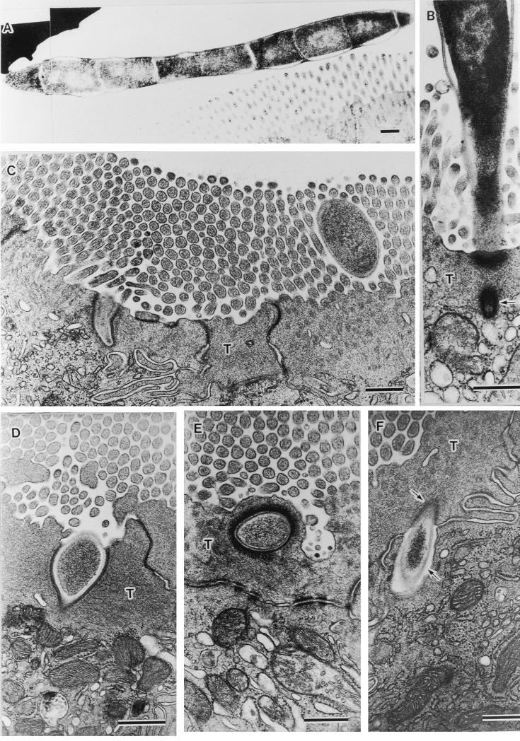
FIG. 2.
Fine structural demonstration of phagocytosis pathway of SFB into ileal epithelial cells. (A) SFB sectioned through its floating part in the intestinal lumen tangentially (magnification, ×11,520). (B) SFB attached to host cell distributing on villus tip, a tip of which is tearing from the SFB body (arrow; ×32,400). C through F, extracellular particles torn on the terminal web (T) (magnifications: C, 23,040; D, 38,250) embedded into T (E: magnification, ×27,000), and engulfed into the cytoplasm beyond T (F: magnification, ×28,800) of host cells distributing on the lower villi. In T, an engulfed particle is surrounded by electron-dense host cytoplasm (upper arrow in F), but in the cytoplasmic area beyond T, it is surrounded by an electron-pale layer (lower arrow in F). Note a similarity in the images of extracellular particles with that of SFB and successive pictures, from the tearing of SFB to its engulfment into host cytoplasm. All bars, 0.5 μm.

