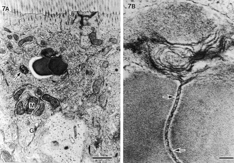
FIG. 7.
Another example of an SFB particle phagocytized into the host cell area beneath the terminal web and cut at the septum. (A) Aggregated mitochondria under the bacteria (M) and well-developed Golgi bodies (G) (scale bar, 1 μm; ×14,400). (B) Higher magnification of an unlacing of septum showing a double-helix-like structure (arrows) and melting at the upper side (scale bar, 0.1 μm; ×108,000).

