Figure 1.
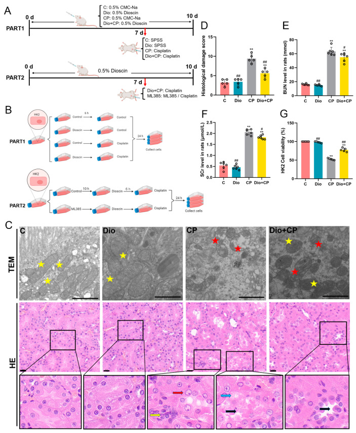
Dioscin relieves cisplatin-induced AKI. (A) Schematic representation of animal study design in the present study. SPSS, stroke-physiological saline solution. (B) Schematic diagram of cell experiment design. (C) Transmission electron microscope (scale bar = 1 μm) and H&E staining (scale bar = 20 μm) of rat kidney tissue (n = 5). Yellow pentagram, normal mitochondria; red pentagram, damaged mitochondria; red arrow, abnormal nucleus; yellow arrow, inflammatory cell infiltrated; blue arrow, renal tubular epithelial cell degeneration; black arrow, renal tubular epithelial cell desquamation. (D) Histological damage score. (E,F) Bun and SCr levels in rat serum (n = 6). (G) HK2 cell activity. C, control group; Dio, dioscin group; CP, cisplatin group; Dio + CP, dioscin + cisplatin group. Results are presented as Mean ± SD. Statistical significance was obtained by one-way ANOVA. ** p < 0.01 compared with C group. # p < 0.05, ## p < 0.01 compared with CP group.

