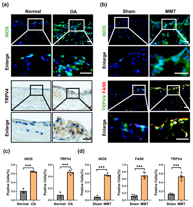
Figure 1.
TRPV4 and M1 macrophages are elevated in OA synovium. (a) Immunofluorescence images of iNOS and immunohistochemical staining of TRPV4 in normal and OA human synovium (Scale bar: 50 μm). (b) Immunofluorescence images of iNOS, TRPV4, and F4/80 in the sham and MMT rat synovium (Scale bar: 50 μm). (c) Percentages of iNOS- and TRPV4-positive cells in human synovium. normal human synovium sample = 3, OA human synovium sample = 6. Unpaired two-tailed t-test, *** p < 0.001. (d) Percentages of iNOS-, F4/80-, and TRPV4-positive cells in sham and MMT group. n = 5. Unpaired two-tailed t-test, *** p < 0.001. All data are shown as mean ± SD.

