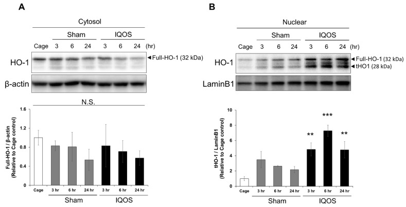
Figure 4.
Alterations in HO-1 protein localization in murine lungs by exposure to IQOS. (A) Expression analysis of HO1 in the cytoplasmic fraction of murine lungs from cage controls, sham controls, and at various times after exposure to IQOS. The density of HO-1 bands was measured, and the expression ratio normalized to β-actin was calculated and expressed as fold-change relative to cage control mice. (B) Expression analysis of nuclear HO-1 in the lungs of at cage controls, sham controls, and at various times after exposure to IQOS. The density of HO-1 bands was measured, and the expression ratio normalized to LaminB1 was calculated and expressed as fold-change relative to cage control mice. ** p < 0.01, and *** p < 0.001 versus cage control group. N.S.: not significant. N = 3. Full-HO-1: full-length HO-1; tHO-1: truncated HO-1.

