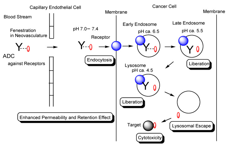
Figure 2.
The pathway of intravenously administered antibody–drug conjugates (ADCs) against receptors such as cancer antigens. ADCs were internalized into cancer cells via receptor-mediated endocytosis (RME). Payloads were liberated by pH-sensitive linker cleavage based on acidification as endosome maturation or by enzymatically cleavable linker cleavage based on lysosomal enzymes and were transported to the cytoplasm by endosomal or lysosomal escape via passive diffusion and/or carrier-mediated transporters. Finally, payloads exhibited anti-cancer activity in the cytoplasm or the nucleus. Y represents a monoclonal antibody (mAb). The blue sphere indicates a receptor that mediates endocytosis in cancer cells. The red ovals represent a drug that is tethered with a mAb through a suitable linker. The black sphere indicates target substances such as topoisomerase II or DNA. The dotted line indicates a linker contained in an ADC. The solid line represents the membrane.

