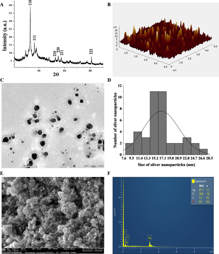
Figure 2.
Characterization of the synthesized silver nanoparticles. (A) XRD showing the crystalline nature of POAgNPs. (B) Atomic force microscopy showing homogeneously shaped silver nanoparticles. (C) TEM images showing spherical-shaped and well-dispersed silver nanoparticles. (D) Histogram of the size distribution of POAgNPs. (E) FESEM micrograph showing the surface topology of silver nanoparticles. (F) Energy dispersive X-ray spectrum of POAgNPs showing the elemental composition of POAgNPs.

