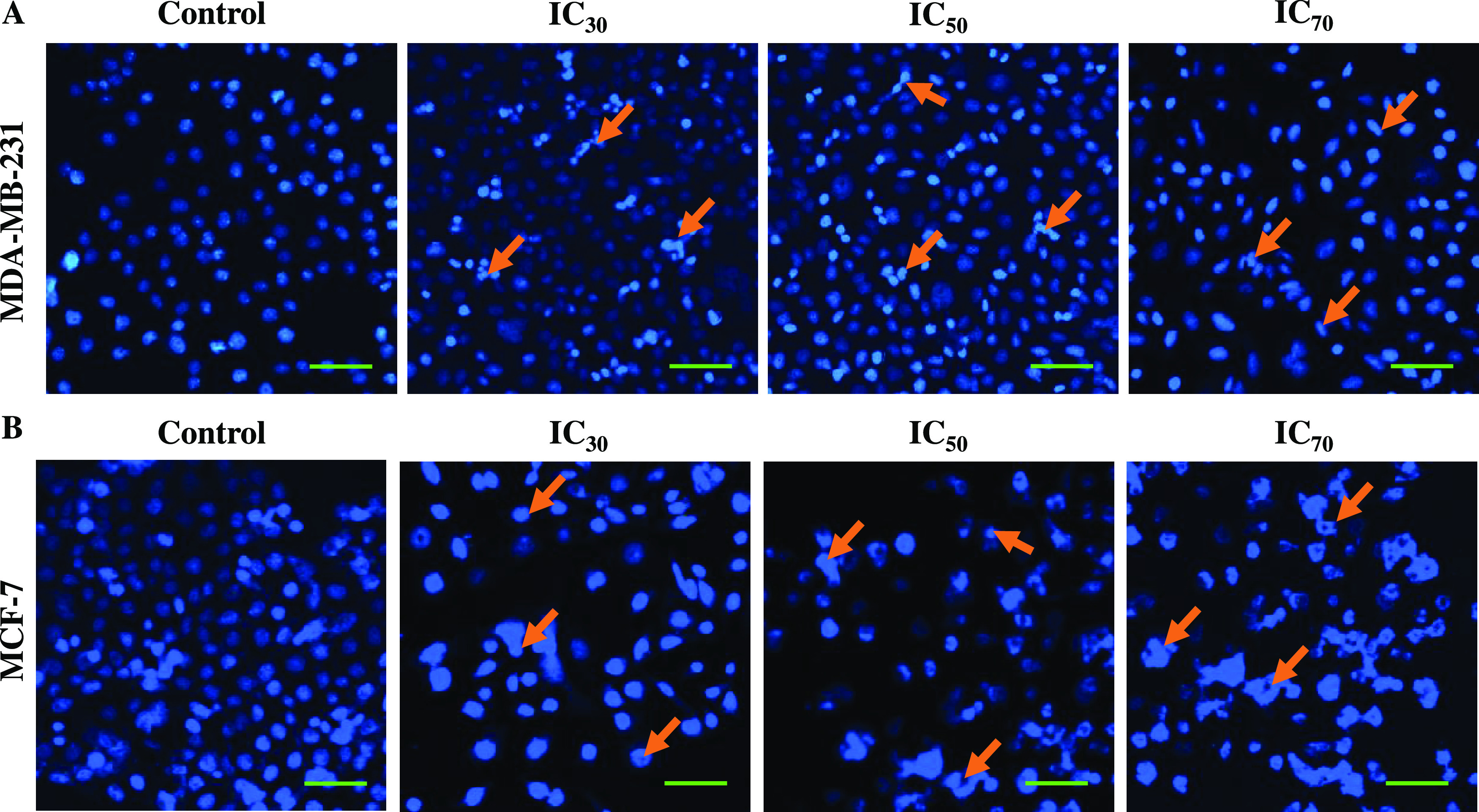
Figure 7.

Fluorescence microscopy images of 4′,6-diamidino-2-phenylindole (DAPI)-stained breast cancer cells treated with POAgNPs in a dose-dependent manner for 24 h. (A) MDA-MB-231 cells. (B) MCF-7 cells. The images represent the morphological changes in the cells treated with IC30, IC50, and IC70 concentrations of POAgNPs as compared to those of control cells. The cells in the control group are observed to have normal rounded nuclei with normal blue color; however, the cells in the treated group are bright in color with condensed chromatin material and abnormal nuclei with an irregular cellular structure (marked through arrows in the figure) that clearly indicates apoptosis of cells. Scale bar = 10 μm.
