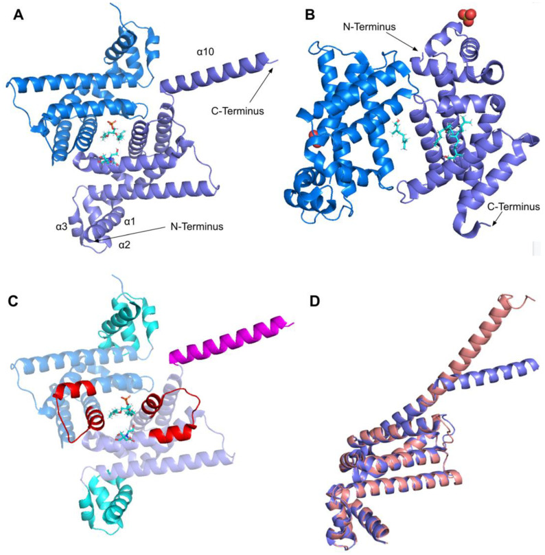
Figure 29.
Comparison of existing structures of COQ9 and the AlphaFold model of COQ9 (A) PDB: 6AWL structure of COQ9ΔN79 with co-crystallized phosphatidylethanolamine and bis-tris shown in cyan, data from [50]. Chain A colored in purple, chain B colored in blue. (B) PDB: 6DEW structure of COQ9ΔN79,ΔC36 with co-crystallized geraniol, cis-trans-farnesol, trans-trans-farnesol, and cis-cis-farnesol shown in cyan and sulfate molecules shown as ball structures; Chain A colored in purple, chain B colored in blue, data from [50]. (C) Structural motifs shown on 6AWL structure. Colors match Figure 28. (D) Structural alignment of 6AWL chain A shown in purple with the AlphaFold model obtained from the AlphaFold Protein Structure Database [39,54], shown in pink.

