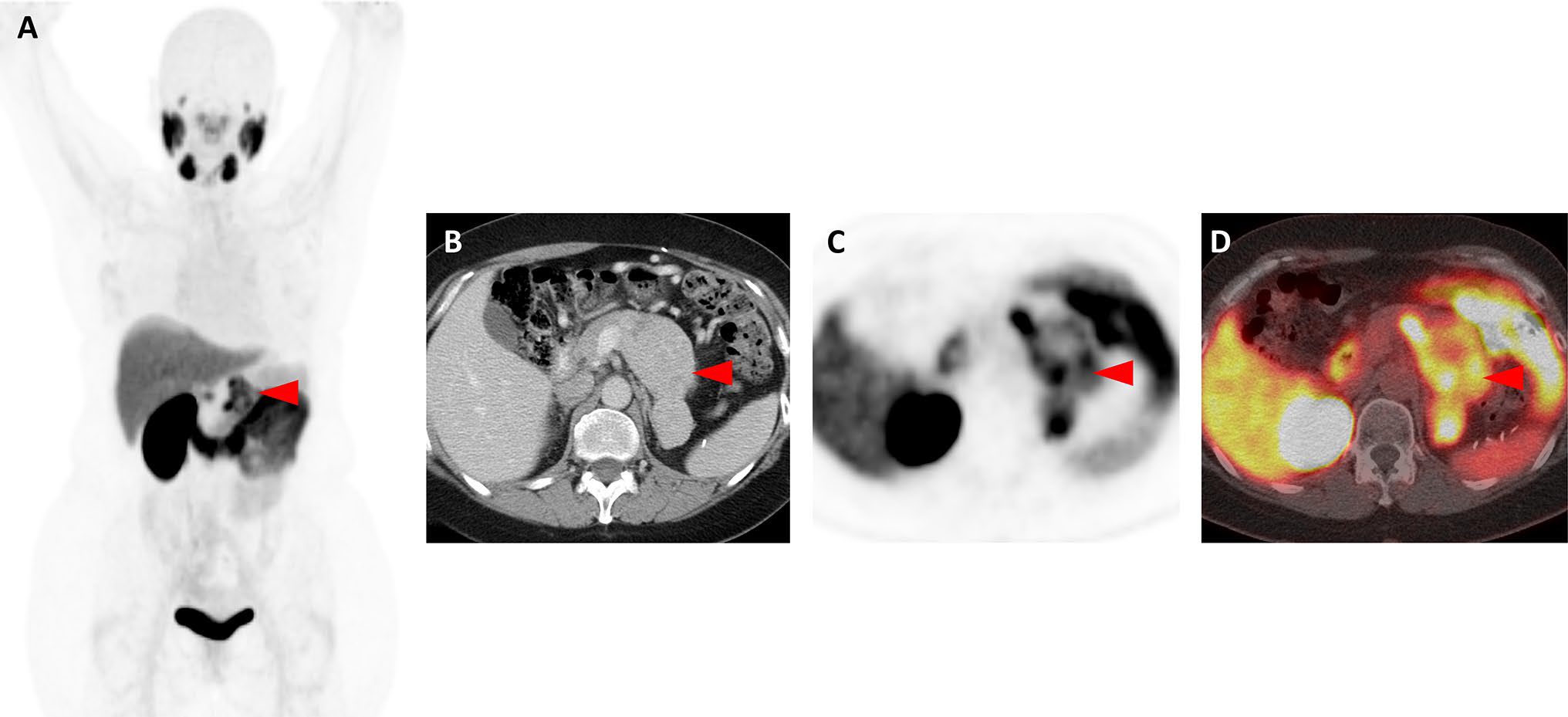
Fig. 1.

Images of a patient with oligometastatic clear cell RCC confirmed with 18F-DCFPyL PET/CT (Patient #12). a Whole-body 18F-DCFPyL PET maximum intensity projection image demonstrates a solitary site of abnormal uptake (red arrowhead) in the region of the left nephrectomy bed. b Axial, contrast-enhanced, venous-phase CT image demonstrates a recurrence in the left nephrectomy bed with tumor invading and expanding the left renal vein (red arrowhead). c Axial 18F-DCFPyL PET and d axial 18F-DCFPyL PET/CT images show focal radiotracer uptake in the lesion (red arrowheads)
