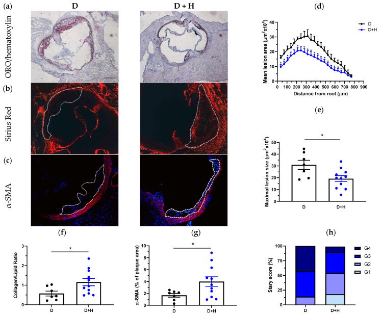
Figure 4.
Hidrosmin treatment reduces atherosclerosis development in diabetic ApoE KO mice. Representative images of ORO/hematoxylin staining (a), Sirius red collagen staining (b) and VSMC immunodetection ((c); red, α-SMA; blue, DAPI nuclear staining) in aortic root sections of diabetic mice untreated (D) and treated with hidrosmin (D + H). Magnification ×100 (a) and ×200 (b,c). (d) Quantification of the extent of atherosclerotic lesions within the aorta. (e) Average of individual maximal lesion size in each group. (f) Assessment of collagen-to-lipid ratio. (g) Quantification of VSMC content in lesions. (h) Classification of mouse atherosclerotic plaques according to the Stary method (grades G1 to G4). Graphs represent individual values and mean ± SEM of each group (D, n = 7; D + H, n = 11). * p < 0.05 vs. D group by Mann–Whitney U test.

