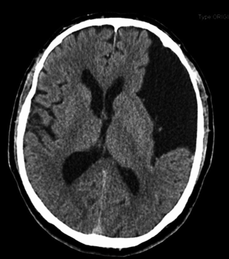
Figure 1. Brain computed tomography.
Brain computed tomography in axial view showing a large left fronto-parieto-temporal extra-axial fluid collection measuring 9.5 x 5.1 cm of maximum diameters creating a significant parenchymal and ventricular compression with subfalcine herniation (midline shift). It is also possible to see a 5 mm deviation of midline structures.

