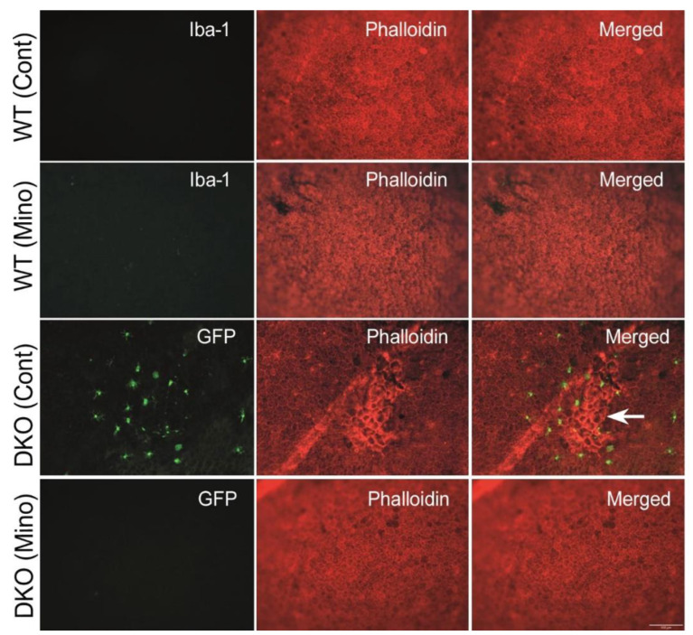
Figure 9.
The effect of minocycline on the structure of retinal pigment epithelial cells. RPE/choroidal flatmounts were stained with phalloidin (red) and Iba-1 (green, for wild-type mice). Microglia in double knockout (DKO) mice were GFP+. Arrow indicating RPE dysmorphia and microglial accumulation. Scale bar = 100 μm. RPE: retinal pigment epithelium.

