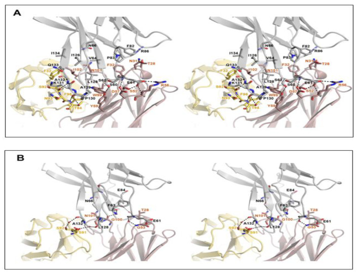
Figure 2.
Detailed interactions within the PD-1/cemiplimab interface. (A) Stereoscopic view of the interactions, including hydrogen bonds, ionic interactions, and van der Waals contacts between PD-1 and cemiplimab. (B) Stereoscopic view of the water-mediated hydrogen bonds. PD-1, heavy, and light chains of cemiplimab are colored gray, pale red, and pale yellow, respectively. Hydrogen bonds and ionic interactions are depicted as dotted lines.

