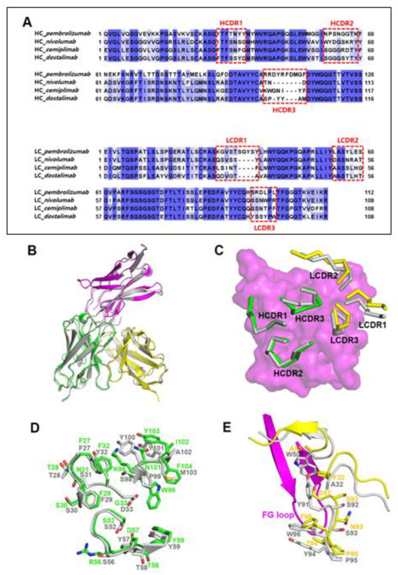
Figure 3.
Similar binding modes of cemiplimab and dostarlimab. (A) Sequence alignments of the variable regions of the FDA-approved anti-PD-1 antibodies. The CDRs are indicated by dotted boxes. HC and LC mean heavy chain and light chain, respectively. (B) Superposition of the PD-1 protein within the cemiplimab/PD-1 and dostarlimab/PD-1 complexes for comparison. The dostarlimab/PD-1 complex is colored gray, and the PD-1, heavy, and light chains in the PD-1/cemiplimab complex are colored purple, green, and yellow, respectively. (C) Conformational comparison of the CDRs within the cemiplimab/PD-1 and dostarlimab/PD-1 complexes. The dostarlimab CDRs are colored gray. HCDRs and LCDRs of cemiplimab are colored green and yellow. PD-1 is represented as a purple surface model. (D) Detailed structural comparison of the residues within the HCDRs of cemiplimab (green) and dostarlimab (gray). (E) Detailed structural comparison of the residues within the LCDRs of cemiplimab (yellow) and dostarlimab (gray). The FG loop of PD-1 in the complex is colored purple.

