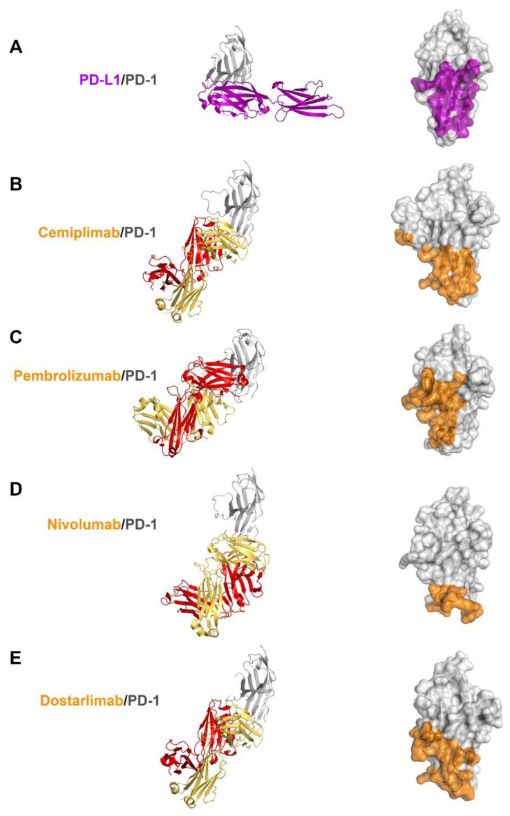
Figure 4.
Comparison of PD-1 binding by PD-L1 and antibodies. (A) Structure of the extracellular domain of PD-1 (gray) in complex with the extracellular PD-L1 (purple) and the PD-L1 binding site (purple) on the surface of PD-1. (B) Structure of the extracellular domain of PD-1 in complex with the cemiplimab Fab and its epitope region on the surface of PD-1. (C) Structure of the extracellular domain of PD-1 in complex with the pembrolizumab Fab and its epitope region on the surface of PD-1. (D) Structure of the extracellular domain of PD-1 in complex with the nivolumab Fab and its epitope region on the surface of PD-1. (E) Structure of the extracellular domain of PD-1 in complex with the dostarlimab Fab and its epitope region on the surface of PD-1. In (A–E), PD-1 is displayed in the same orientation, and the heavy and light chains of the antibodies and their epitope regions are colored red, yellow, and orange, respectively.

