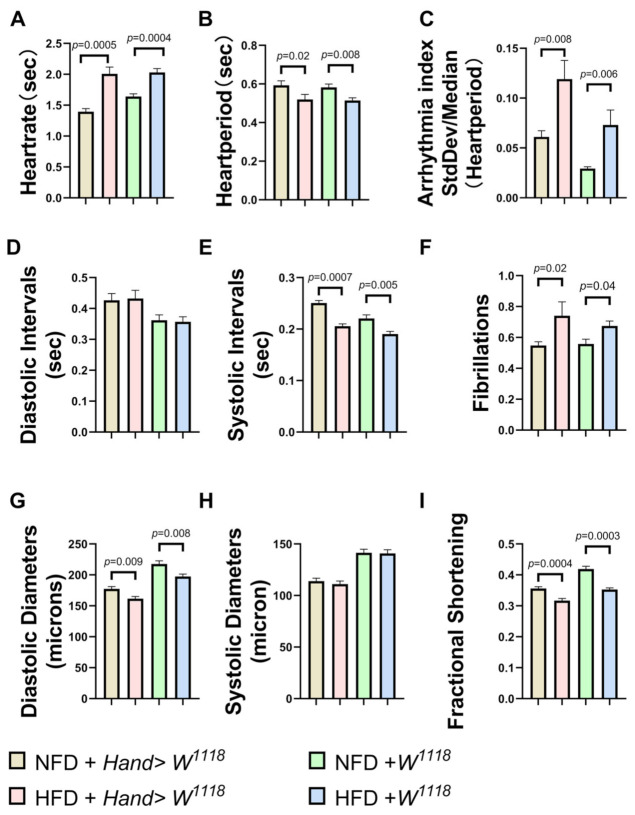
Figure 3.
NFD and HFD groups of 10-day-old Drosophila M-mode ECG, with quantitative analysis of HR, HP, AI, DI, SI, FL, SD, DD, FS. Compared to NFD, w1118 and Hand > w1118 under HFD conditions, HR, AI and FL (A,C,F) were significantly upregulated. Moreover, HP, SI, DD and FS (B,E,G,I) decreased significantly, while DI and SD (D,H) did not show significant differences. All Drosophila were virgin flies, and the sample sizes for the two groups of NFD and HFD were N = 28 and N = 30, respectively. All p values were from independent samples t-tests.

