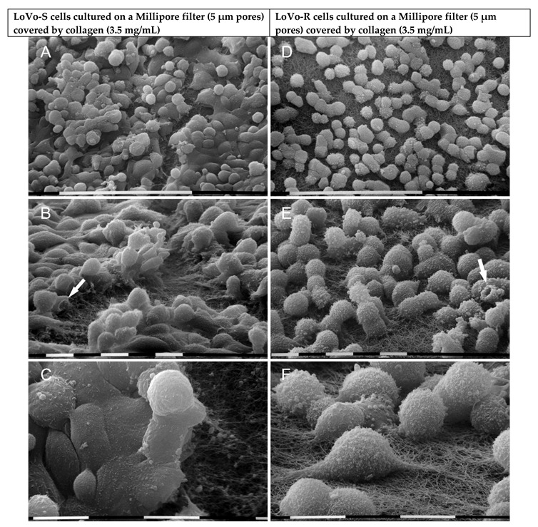
Figure 6.

Ultrastructural morphological characteristics of LoVo-S and LoVo-R cells cultured for 24 h on Millipore/type I collagen mimicking a desmoplastic lamina propria. LoVo-S cells include polygonal cells in tight cell-cell contact with each other and adhering to the collagen fibrils, which are only partially visible. However, globular-shaped cells grow on the flattened ones (A,B). A funnel-shaped cell is detectable (arrow) (B). LoVo-S cells show few microvilli, but exosomes and microvesicles are embedded in the collagen fibril network (C). The LoVo-R cells look like globular-shaped cells next to each other, but no tight contacts are visible, so collagen fibrils are always distinguishable (D). A funnel-shaped cell is detectable (arrow) (E). The LoVo-R cells develop many microvesicles on their surface and appear strongly adherent to the fibrils with also filopodia invaginating the micropores of the collagen fibril network (E,F). Bar = 100 µm (A,D) Bar = 10 µm (B,C,E,F).
