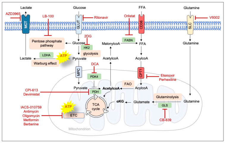
Figure 3.
The cell’s main metabolic pathways and the corresponding inhibitors. Glucose, free fatty acids (FFAs) and glutamine enter cells via glucose transporters (GLUT), CD36, and solute carrier (SLC) transporters respectively. Once inside the cells, these substrates are catabolized by various metabolic reactions in the cytosol and the mitochondria in the tricarboxylic acid cycle (TCA). Cofactors produced by the TCA are used by the electron transport chain (ETC) to produce ATP. Some cancer cells shift their metabolism (and specifically glycolysis) towards lactate production; ATP is generated outside the mitochondria. Lactate can then be exported from the cells via monocarboxylate transporters (MCTs). Inhibitors are shown in red, key enzymes are labeled in green, and metabolic pathways are shown in pink. αKG, α-ketoglutaric acid; FASN, fatty acid synthase; FAO, fatty acid oxidation; GLS, glutaminase; HK2, hexokinase 2; LDHA, lactate dehydrogenase A; PDH, pyruvate dehydrogenase; PDK4, pyruvate dehydrogenase kinase 4.

