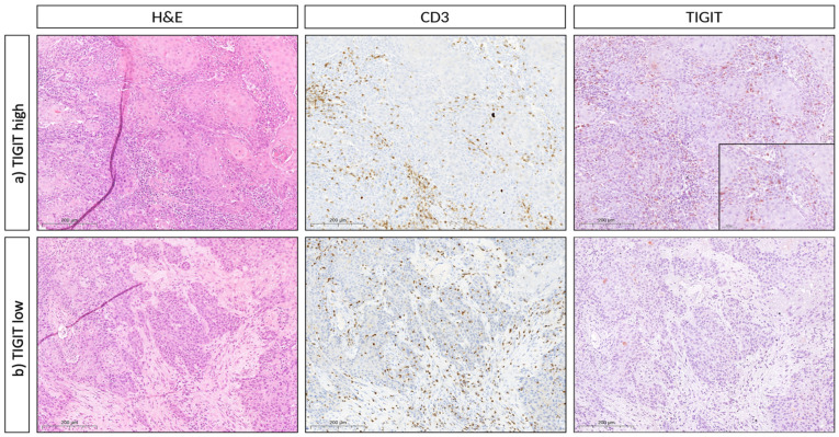
Figure 3.
Immunohistochemical TIGIT expression in OSCC TMA cohort. Representative histopathology (hematoxylin & eosin, H&E) and CD3/TIGIT immunohistochemistry (IHC), respectively, of two cases of oral squamous cell carcinoma (OSCC). (a) (First row) represents an OSCC with stromal and intraepithelial CD3 and TIGIT double-positive T cells (magnification 400X each, 1000X for the inset). (b) (Second row) represents an OSCC with stromal and intraepithelial CD3+ T cells that do not express TIGIT (400X).

