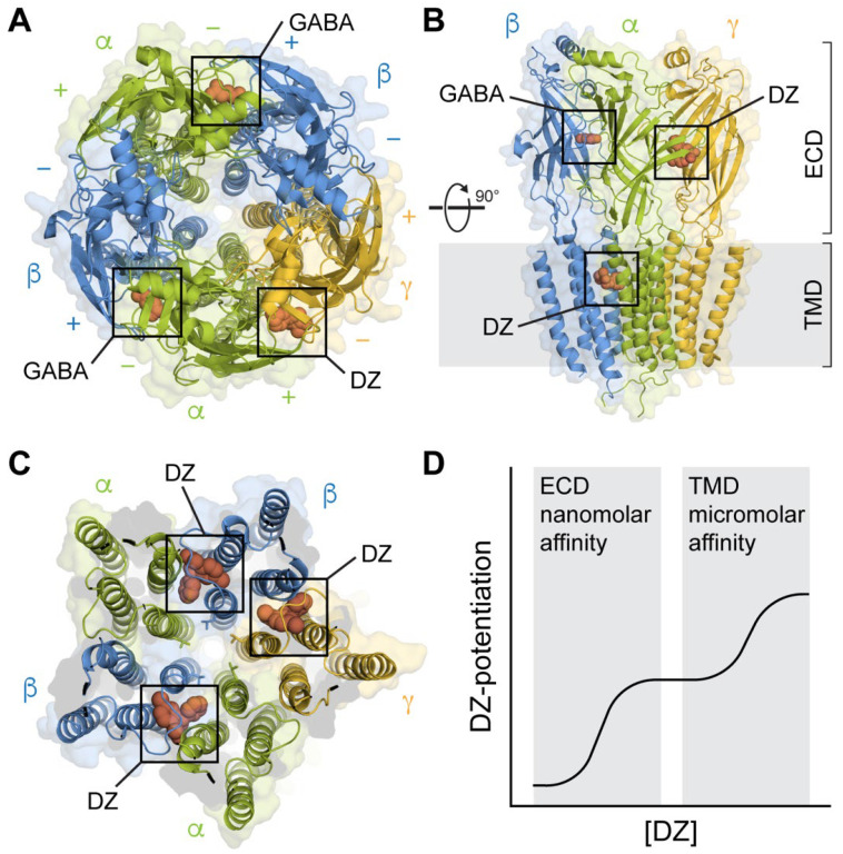
Figure 1.
Structures of synaptic GABAARs in complex with the BZD PAM diazepam (DZ) at both high-affinity ECD and lower-affinity TMD sites. (A) Top-down view with GABA bound at the β+/α− interfaces and DZ bound at the α+/γ− interface in the ECD. Cryo-EM map of GABAA(α1)2(β3)2(γ2)1 from PDB 6HUP. (B) Side view of the structure in A, additionally showing one of several binding sites for DZ in the TMD. Only three subunits are shown for clarity. (C) Same perspective as in (A) for a slice through the TMD. Cryo-EM map of GABAA(α1)2(β2)2(γ2)1 from PDB 6X3X. Three binding sites for DZ are highlighted at the β+/α− and γ+/β− intersubunit pockets between the TM helices and below the M2-M3 linker of one of the subunits. The central pore-gate-forming 9′ leucine residues are shown as sticks near the middle of the pore-lining M2 helices. (D) Biphasic modulation by DZ at the canonical high-affinity site in the ECD and lower-affinity sites in the TMD.

