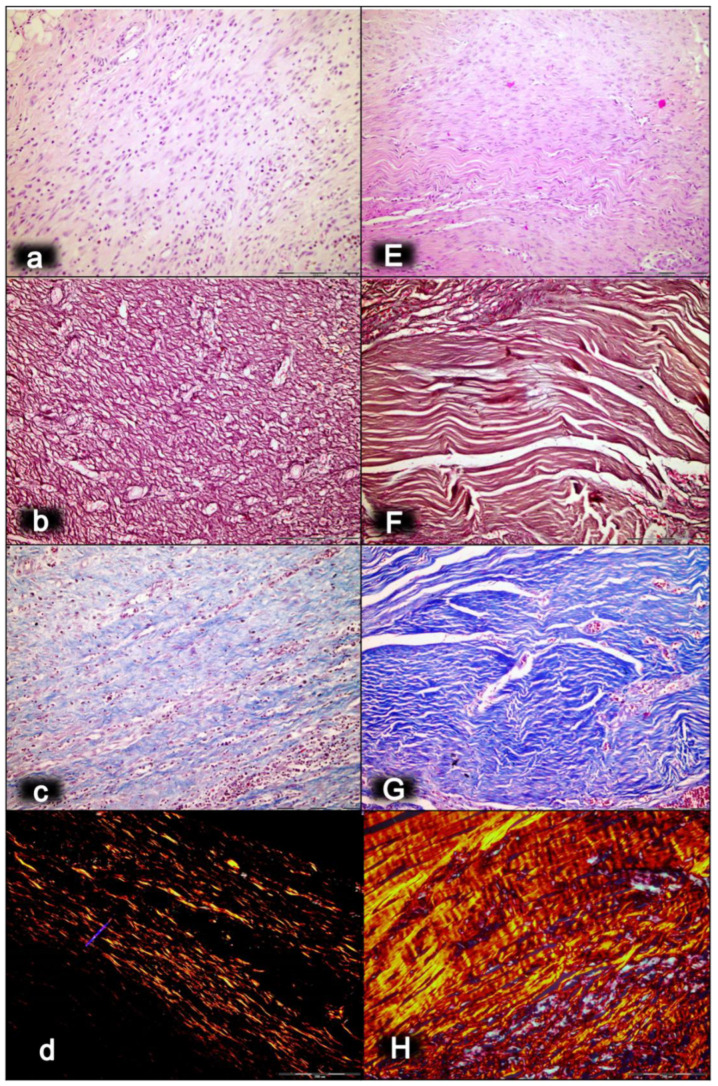
Figure 3.
Myotendinous junction 28 days following dissection of quadriceps tendon from the quadriceps muscle [43]. Regular presentation after myotendinous junction injury induction in control rats (small letters) (a–d). No edema and inflammatory cells were found with vanishing myotendinous junction revascularization (a). The proliferation of fibroblasts and fibers, both reticulin (b) and collagen fibers (c), as well as fiber maturation (much less than in BPC 157 treated rats) with suboptimal orientation to the long axes of the myofibers close to the myotendinous junction. Regular presentation after myotendinous junction injury during BPC 157 regimen (capitals), given in drinking water (E–H). Well-oriented dense connective tissue was found (E). Morphologic features of the myotendinous junction area indicate that BPC 157 therapy favors vascular density as well as reconstruction and orientation of reticulin (F) and collagen fibers (G). A prominent proliferation of fibroblasts and fibers as well as fiber maturation was obtained (confirmed with polarized microscope; H), providing tenable fibroblast and fiber proliferation with optimal orientation due to the long axes of the myofibers close to the myotendinous junction with a lesser number of fibroblasts, and with a higher amount of reticulin and collagen fibers. In contrast, the control group showed far less abundant and well-oriented collagen type 1 fibers (d). HE staining (a,E); histochemical Gomori staining (b,F) and Masson trichrome staining (c,G); Sirius red histochemical staining with polarized microscopy (d,H); magnification ×200.

