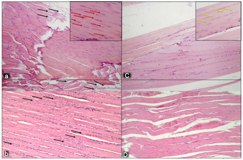
Figure 5.
Myotendinous junction six months following dissection of quadriceps tendon from the quadriceps muscle [43]. Regular presentation after myotendinous junction injury induction in control rats (small letters) (a,b). Tendons have significant numbers of lipid-like structures at the myotendinous junction wound site (black arrows) (a). Regular presentation after myotendinous junction injury during BPC 157 regimen (capitals), given in drinking water (C,D). No lipid-like structures appeared in treated animals (C). Also, there is a significantly increased number of typical tendon cells that are more elongated in the treated animals (yellow arrows) (C), while there is an increased number of round shaped cells, presumably non-tenocytes, in the control group (red arrows) (a). The myotendinous junction is at a high risk for strain injuries, due to the high amounts of energy that are transferred through this structure, indicating the remodeling capacity of the myotendinous junction. The general feature found within a peak period of remodeling is a progressive restoration of the tissue by neosynthesized fibers that are centronucleated. We observed the degree of remodeling of muscle fibers alike the human myotendinous junction in the control group (b), where a high portion of the muscle fibers adjacent to the myotendinous junction contained a centrally located myonucleus. This is suggestive of a very high rate of remodeling of the muscle fibers near the myotendinous junction in control group (black arrows) (b) in comparison with treated animals (D). There were notable differences in the cell kinetics remodeling of fibrils at the myotendinous junction between control and BPC 157 group. HE staining; magnification ×200.

