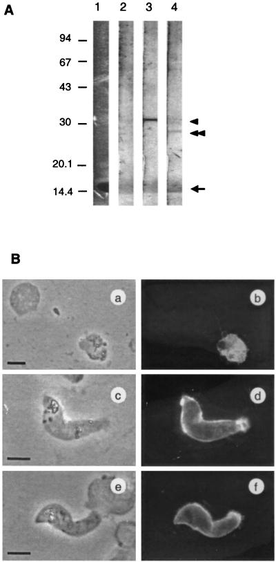
FIG. 5.
Immunological reactivity of antisera to clinically isolated P. vivax. (A) Western blotting analysis of cultured P. vivax. Protein extracts from cultured parasites isolated from patients were size fractionated by SDS-PAGE under nonreducing conditions, and proteins were transferred to a polyvinylidene difluoride filter. Filter strips were incubated with anti-PfMSP-119 (lane 2), anti-Pvs25 (lane 3), or anti-Pvs28 (lane 4) mouse antiserum. Pvs25 (arrowhead) or Pvs28 (double arrowhead) were stained by antiserum against Pvs25 or Pvs28. Nonspecific staining was observed at around 15 kDa (arrow) coincident with an abundance of protein as determined by Coomassie blue staining (lane 1). Positions of standard molecular size markers are indicated in kilodaltons on the left. (B) IFA of cultured P. vivax sexual-stage parasites. Ice-acetone-fixed parasites were stained with anti-Pvs25 (a to d) or anti-Pvs28 (e and f) mouse antiserum from CAF1 mice. IFA (a, c, and e) and phase-contrast (b, d, and f) images of identical fields are shown. Bars = 5 μm.

