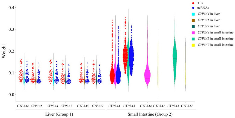
Figure 5.
Comparison of CYP3A-associated TFs and ncRNAs with three CYP3As in the liver and small intestine. The y-axis is for the weight values calculated by WGCNA. Red and blue dots, respectively, represent the overlapping TFs and ncRNAs associated with two different CYP3As in the liver or small intestine. The violin plots with same color represent the same CYP3A in the same tissue.

