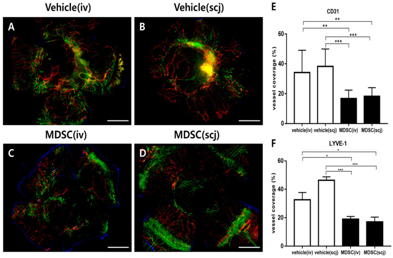
Figure 5.
Comparisons of neovascularization and lymphangiogenesis in corneal allografts via MDSC administration. Representative whole-mount corneal immunofluorescent CD31hi (green) and LYVE-1hi (red) staining images from each group: PBSiv (A), MDSCiv (C), PBSscj (B), and MDSCscj (D). The representative photograph is a combination of multiple stitched photographs (5× magnification, scale bar = 500 μm) taken after dividing the whole cornea into several parts. MDSC groups (MDSCiv, MDSCscj) presented the significantly suppressed areas of neovascularization (E), green-CD31hi; white arrows for blood vessels and lymphangiogenesis ((F), red-LYVE-1hi) rather than those of the vehicle groups (PBSiv, PBSscj). Data are presented as the mean ± SEM of three repeated experiments involving three corneas per group (E,F); * p < 0.05, ** p < 0.01, and *** p < 0.001.

