Figure 1.
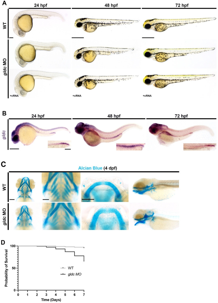
A severe gldc deficient model exhibits phenotypes consistent with impaired kidney function. (A) Live images of WT and gldc morphants at 24 hpf, 48 hpf, and 72 hpf. Images at 24 hpf reveal gray pallor within the head of gldc morphants. At 48 hpf, pericardial edema becomes evident and persists through 72 hpf. At 72 hpf, craniofacial cartilage begins to develop, and abnormalities are observed. Scale bars = 100 μM, 400 μM. (B) WISH of WT gldc expression at 24 hpf, 48 hpf, and 72 hpf reveals gldc transcripts within the CNS and pronephros. Scale bars = 200 μM (main image) and 50 μM (inset). (C) Alcian Blue cartilage staining in WT and gldc morphants at 4 dpf. Decreased number of pharyngeal arches and aberrant jaw morphology were seen in gldc morphants. Scale bars = 100 μM, 50 μM, 50 μM. (D) A survival curve of WT animals and gldc morphants reveals that gldc deficient animals have a decreased percent survivability over seven days.

