Figure 3.
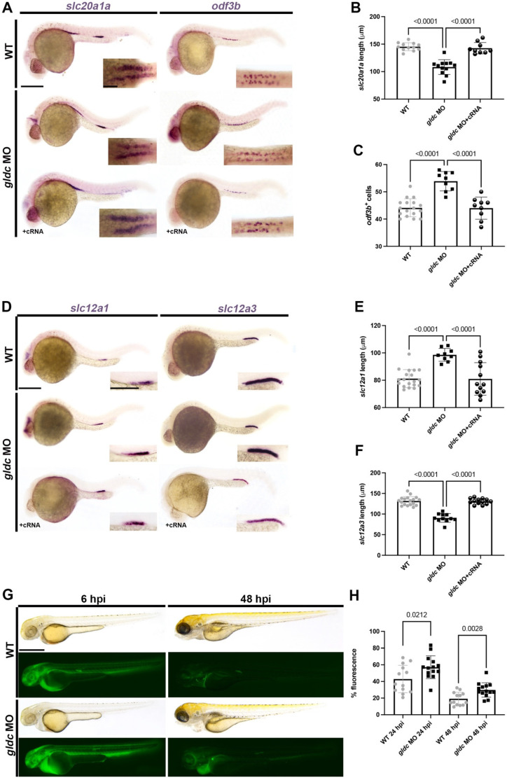
gldc is necessary for proper segment patterning. (A) WISH of slc20a1a and odf3b in WT, gldc MO, and gldc MO + cRNA at 24 hpf. Scale bars = 200 μM (main image) and 50 μM (inset). (B) Absolute length quantification of slc20a1a domain in control and treatment groups. (C) Quantification of odf3b+ cells in WT, gldc MO, and gldc MO + cRNA. (D) WISH of slc12a1 and slc12a3 in WT, gldc MO, and gldc MO + cRNA at 24 hpf. Scale bars = 200 μM (main image) and 50 μM (inset). (E,F) Absolute length quantification of slc12a1 and slc12a3 domains, respectively. (G) Animals were injected with dextran-FITC at 24 hpf, then imaged at 6 hpi and 48 hpi. Scale bar = 400 μM. (H) Quantifications of percent fluorescence at 24 hpi and 48 hpi. Percent fluorescence was calculated with 6 hpi fluorescent intensity as baseline. Data are mean ± s.d. quantified for each control and experimental group. Absolute lengths and odf3b+ cell counts were compared using ANOVA. Percent fluorescence measurements were compared with unpaired T-tests.

