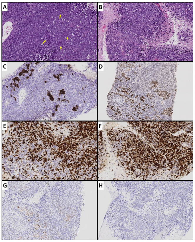
Figure 3.

Metastatic de-differentiated carcinoma in a patient with well-differentiated pancreatic neuroendocrine tumor. (A) Representative tumor section containing foci of well-differentiated component (yellow arrows). (B) Poorly differentiated carcinoma with extensive necrosis and abundant apoptosis and mitoses. (C) Well-differentiated foci with synaptophysin expression but no expression in poorly differentiated component. (D) Well-differentiated foci with strong INSM-1 (another neuroendocrine marker) expression. (E,F) Relatively low Ki67 index in well-differentiated component and high Ki67 poorly differentiated component. (G) Normal p53 expression in well-differentiated component. (H) Complete loss of p53.
