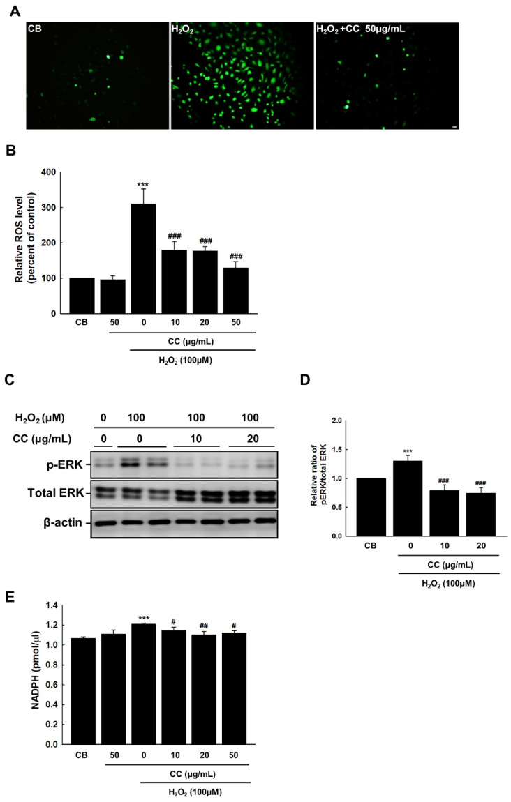
Figure 2.
Chrysanthemum coronarium L. (CC) extract reduces endothelial senescence in HUVECs induced by ROS. (A) Fluorescent staining images illustrating ROS production in HUVECs via DCF-DA staining. The relative ratios of DCF-DA fluorescence intensities indicate the degree of endothelial ROS formation in HUVECs. The original magnification was set to 20 ×. Scale bar = 50 μm. (B) The fluorescence sensitivity of DCF-DA was standardized for the total number of cells in each dish. (C–D) Expression of phospho-ERK, total ERK, and β-actin in H2O2-treated cells. (E) NADPH was measured using a colorimetric assay. Control cells (CB) received the vehicle alone. Bars represent the mean ± SEM from three dishes per group. ***, p < 0.001, versus CB; #, p < 0.05, ##, p < 0.01, ###, p < 0.001, vs. H2O2.

