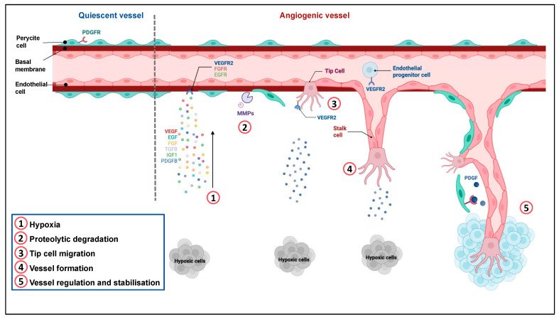
Figure 1.
Regulation of angiogenesis. Quiescent ECs form a thin layer of single flat cells that line the interior surface of blood vessels and lymphatic vessels. These cells are interconnected by junctional molecules. EC monolayer is covered by pericytes, which control EC proliferation, release cell-survival signals and produce the basement membrane. ①. Hypoxia induces the secretion of pro-angiogenic factors. ②. This secretion leads to pericyte detachment, basement membrane degradation by metalloproteases and loss of EC junctions via VEGFR-2 activation. ③. One EC called tip cell is selected to guide elongation of the vessel towards proangiogenic signals. Remodeling of the existing matrix allows the migration of ECs. ④. Stalk cells proliferate and elongate to form a new vessel. ⑤. Following Dll4-Notch signaling and PDGF release, EC resume their quiescent state, the vessel is stabilized by recruitment of pericytes via PDGFR and deposition of a basement membrane. Adapted from “Tumor vascularization”, by BioRender.com (2022). Retrieved from https://app.biorender.com/biorender-templates (accessed on 4 December 2022).

