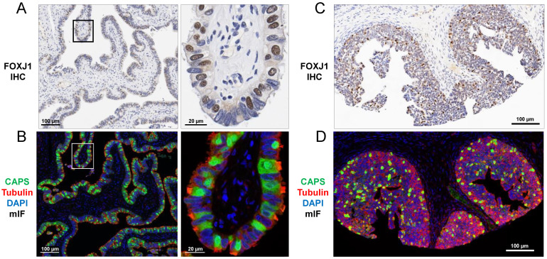
Figure 1.
Ciliated cell markers FOXJ1 (IHC) and CAPS (mIF) in (A,B) normal fallopian tube and (C,D) proliferative endometrium. FOXJ1 shows nuclear staining (A,C) while CAPS shows diffuse intracellular staining (B,D). TUBB4 stains the cytoskeleton including base of cilia (B,D). Images (left) are shown at higher magnification (right). (A,C) Hematoxylin counterstain. (B,D) DAPI counterstain.

