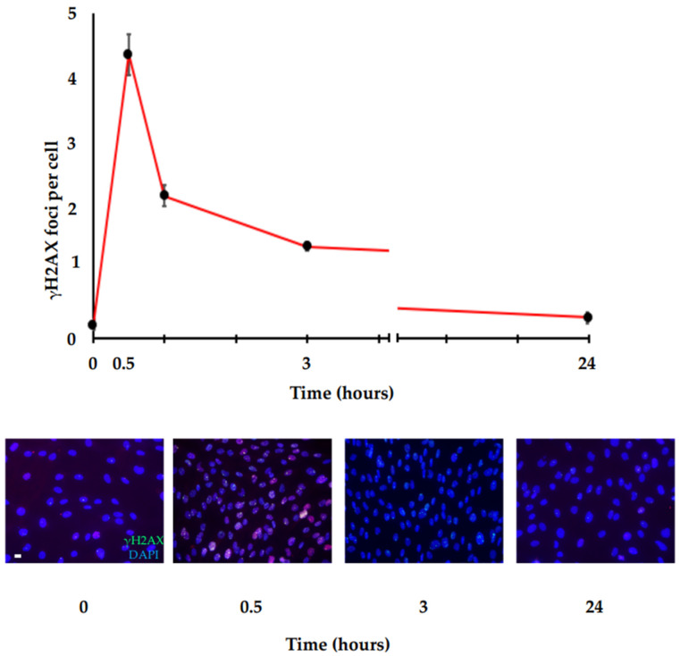
Figure 3.
γH2AX foci repair kinetic. Immunofluorescence analysis of γH2AX in HF cells irradiated with 0.2 Gy. Following DNA damage, γH2AX is quickly induced and reaches the maximum peak in a very short time (about 30 min). Then, the γH2AX levels decrease more slowly until they reach the basal levels at about 24 h. Top panel: graph showing the amount of γH2AX foci/cells at the indicated time points after IR stimulus. Bottom panel: representative immunofluorescence images at the indicated time points after IR stimulus (scale bar is 10 µm).

