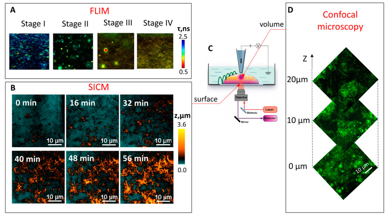
Figure 1.
(A) FLIM images of the Fmoc-FF hydrogel formation at different stages in the presence of 40 µM ThT. Image size 80 × 80 µm2. (B) The process of Fmoc-FF self-assembly as revealed by SICM. (C) Schematic representation of areas in the sample from which by SICM and confocal microscopy images are obtained (D) Confocal microscopy Z-stack images of peptide Fmoc-FF aggregates.

