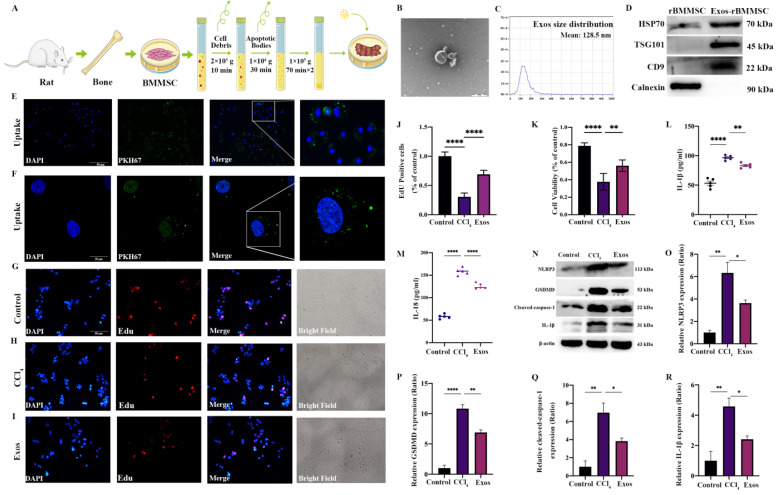
Figure 3.
Effects of Exos-rBMMSCs on CCl4-induced hepatocytes. The extraction protocol of Exo-rBMMSCs is shown (A). TEM morphology and nanoparticle tracking analysis (NTA) results are shown (B,C). HSP70, TSG101, CD9 and Calnexin expression in the rBMMSC and Exos-rBMMSC groups is shown (D). PKH67-labelled Exo-rBMMSC were internalized by hepatocytes, as observed under a fluorescence microscope (E,F). Hepatocyte proliferation in the Exos group was dramatically increased ((G–K), ** p < 0.01 vs. the CCl4 group). IL-1β expression in the Exos group was significantly decreased ((L), ** p < 0.01 vs. the CCl4 group), and IL-18 expression in the Exos group was notably decreased compared to the CCl4 group ((M), **** p < 0.0001). NLRP3, GSDMD, cleaved-caspase-1 and IL-1β expression in the Exos group was significantly decreased compared to that in the CCl4 group ((N–R), * p < 0.05). The data are displayed as the means ± SD; n = 5 for each group.

