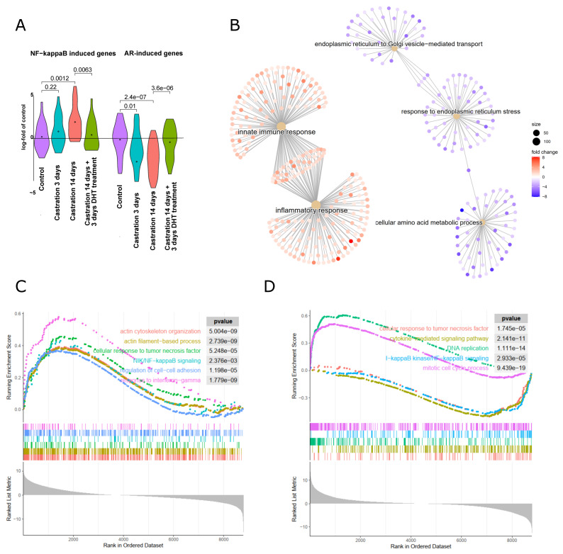
Figure 6.
Gene expression changes in the mouse model of antiandrogen treatment via castration. (A) Gene expression data from prostates derived from mice that were castrated to eliminate androgen signaling, or sham-operated (control) followed by RNA extraction after 3 days or 14 days as indicated. One cohort was treated with dihydrotestosterone for 3 days, 14 days after castration to reactivate androgen signaling (castr. 14 d + DHT 3 d, dataset GEO GSE5901, [28]). Androgen-induced genes [46] and NF-κB target genes (Supplementary Table S1) are shown as violin plots in comparison to control prostates. (B) Comparison of biological processes in prostates of mice 3 days after castration as compared to sham-operated mice: gene ontology analysis of upregulated (red nodes) or downregulated (blue nodes) processes. (C) Gene set enrichment analysis (GSEA) of mice 14 days after castration as compared to sham-operated control mice. Inflammatory signaling pathways, cell adhesion, and actin-based processes are upregulated. (D) GSEA after readdition of testosterone to castrated mice as compared to the castrated mice: mitotic cell cycle and DNA replication are upregulated and NF-κB signaling processes (response to TNFα, cytokine-mediated signaling, and IKK/NF-κB signaling) are downregulated.

