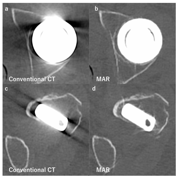
Figure 4.
Comparison of conventional computed tomography (CT) and images reconstructed with the metal artifact reduction algorithm (MAR). Axial images of a patient after hemiarthroplasty of the hip. Images (a,b) show the bipolar femoral head, while (c,d) show the femoral stem at the level of the minor trochanter. Images (a,c) were obtained by conventional CT, and show strong beam hardening, scattering, photon starvation, and edge effects. On the other hand, there are no obvious metal artifacts on images (b,d) using MAR. It leads to better visualization of the outlines of the prosthesis.

