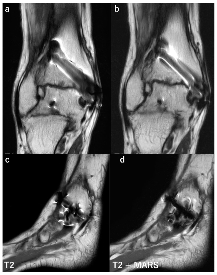
Figure 6.
Comparison of conventional MRI and images obtained with MARS. Coronal and sagittal MR images of a patient after medial malleolus fracture of the right ankle. The fracture was treated by a screw fixation. The coronal MR image (a) was taken by conventional T2 weighted image (T2WI), and significant signal loss and distortion around the implant are present. Image (b) was taken by T2WI with MARS, showing a better visualization of tissue around the screw. The sagittal image (c,d) was taken by T2WI, and T2WI with MARS, respectively. Similarly, with coronal images, signal loss and distortion are reduced with MARS, leading to better visualization of tissue surrounding the implant.

