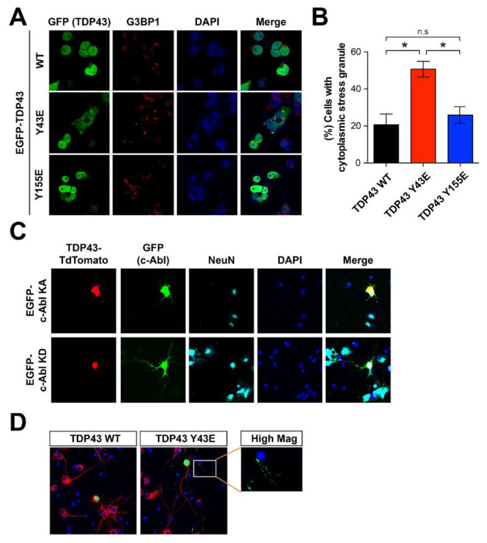
Figure 4.
Y43 residue of TDP-43 associates with the TDP-43 pathology. (A) Immunostaining with G3BP1 antibody that can detect cytoplasmic stress granules in SH-SY5Y cells transfected with GFP-WT TDP-43, GFP-phospho-mimic forms of Y43E and Y155E TDP-43. (B) Quantification of the percentage of cells with cytoplasmic stress granules (n = 3). Statistical significance was determined by using one-way ANOVA with Tukey’s correction; * p < 0.05, n.s. = non-significance. (C) Increased localization of TDP-43 at neurite granules by c-Abl KA co-expression in cultured cortical neurons. TDP-43 TdTomato and EGFP-c-Abl were expressed in the cultured cortical neurons and followed by immunocytochemistry. (D) Phospho-mimic form of TDP-43 Y43E showed increased neurite granule expression than TDP-43 WT in primary cultured cortical neurons. EGFP-TDP-43 WT and Y43E were expressed alone in the cultured cortical neurons and followed by immunocytochemical alayses.

