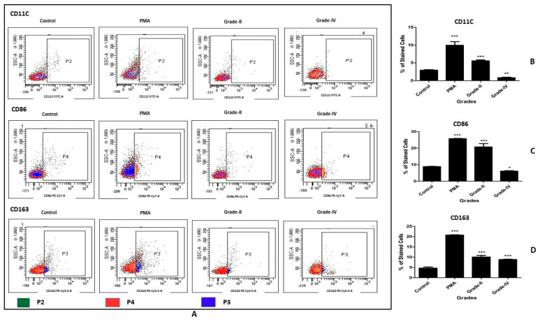
Figure 4.
In vitro cell differentiation assay on PBMC-derived CD14+ cells. PBMC-derived CD14+ cells were induced with SF of KL grade II and IV for 48 h; the status of the newly differentiated cells was evaluated with the cell surface markers CD11C, CD86, and CD163. (A) shows representative scatterplots of the differentiated macrophages and their subsets; CD11C and CD86 are M1 markers, while CD163 is an M2 subtype marker; the gating was based on isotype control. (B–D) shows a grade-wise staining pattern of CD11C+, CD86+, and CD163+ cells; to create these bar graphs, the average percentage of each cell surface marker estimated from three SF samples of each KL grade was used (n = 6); the experiment was performed in triplicate and repeated three times. * p < 0.05, ** p < 0.01, *** p < 0.001, as compared to control.

