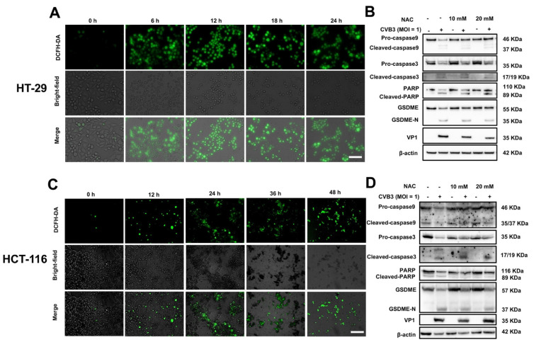
Figure 4.
CVB3-induced pyroptosis is facilitated by ROS. (A,C) Brightfield and fluorescence images of CVB3-infected cells. ROS concentration in HT-29 and HCT-116 cells was measured using DCFH-HA, with green fluorescence indicating the presence of ROS. Magnification, ×100. Scale bar, 100 μm. (B,D) HT-29 and HCT-116 cells were pretreated with or without NAC (10 or 20 mM) for 2 h and then infected with CVB3 for 24 h, and cleavage of caspase-3 and GSDME was analyzed using Western blots.

