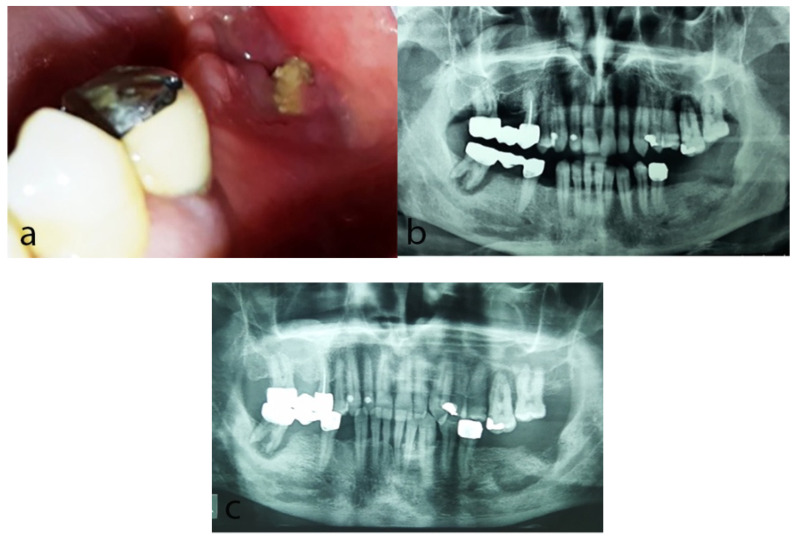
Figure 2.
Case 2: (a) Clinical examination at the first visit revealing exposed necrotic bone in the left retromolar area at the site of a previous extraction before 8 months. (b) Panoramic radiograph at the first visit showing diffuse radiolucency in the left retromolar area. (c) Panoramic radiograph 9 months later showing persistence of the radiolucency and bone sequestrum formation in the same area.

